A Non-Invasive Method Developed to Assess Burn Wound Healing

A Non-Invasive Method Developed to Assess Burn Wound Healing

  • Research Stash
  • News
  • 3.5K

Assessing a burn wound to know the status of healing is currently an invasive process such as biopsy which is both painful and scarring. Now scientists have developed a non-invasive technique that involves mere flash of laser light to assess healing.

The method exploits a particular property of some tissue proteins – ability to re-emit light upon absorption. Such proteins, known as tissue fluorophores, have chemical compounds that can re-emit light. Collagen is one such protein that is vital in wound healing. So when laser light is flashed on tissues under examination, the amount of re-emitted light from the healing tissue directly corresponds to collagen concentration, which in turn, indicates the status of the recovery process.

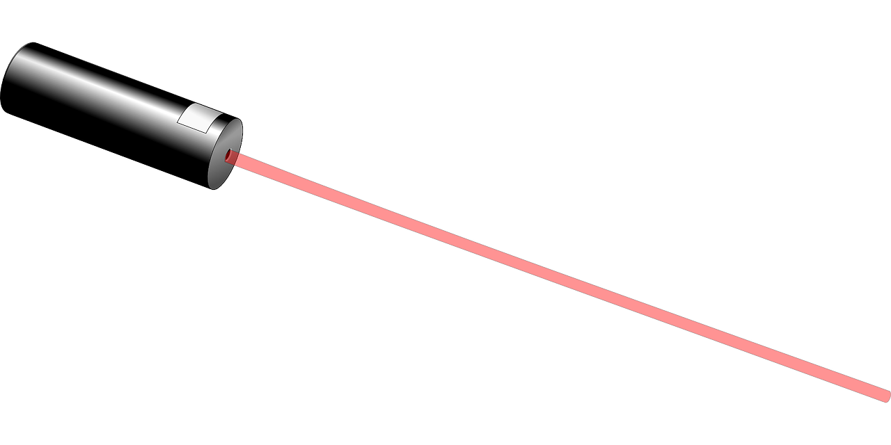
Researchers at the Manipal Academy of Higher Education used commercially available 325 nm laser light to inspect healing by exploring tissue fluorophores, and a detector to collect re-emitted light. The re-emitted spectra were recorded using a fiber optic probe kept very close – about three mm – to the wound site. The spectra were then analyzed in a spectrograph connected to a computer.

The collagen levels measured by this technique, known as autofluorescence, were validated by biochemical tests of patient tissues. The comparison showed that autofluorescence was consistent and suitable for the assessment of burn wound healing. This study also shown tissue collagen can be used as an optical biomarker for assessing burn wound healing, researchers explained. Tissue samples were collected from burn patients admitted to the Department of Plastic Surgery and Burns, Kasturba Medical College.

In earlier studies, the technique was used in lab-induced wounds in animals. Now it has been tested in tissue samples from human burn patients suffering from a different degree of burn wounds. Tissue formed following treatment was excised before grafting and utilized for autofluorescence measurement.

“Our technique is user-friendly and solely dependent on excitation and emission of tissue fluorophores without the addition of any external dye molecule. This helps maintain tissue architecture during the assessment of healing,” Dr. Vijendra Prabhu of Department of Biotechnology at the Manipal Institute of Technology, a member of the research team, told India Science Wire.

For clinical management of wounds, periodic evaluation of injured tissue is necessary to know the progress of healing. Sometimes wound assessment is done by trained clinicians but it is a subjective method. The other method is to conduct histopathology and biochemical analysis to measure the amount of collagen deposited. Repetitive invasive tests can result in fresh wounds and the possibility of infection and scarring.

Through autofluorescence technique, collagen information of the tissue can be gathered just 10 to 15 seconds. “It can provide complementary data conducive to making clinical decisions. After successful testing on clinical samples, we are ready for mass testing on burn wound patients. This system can also be utilized to test collagen disorders in patients,” Dr. Prabhu said.

The research team included researchers from Manipal Academy of Higher Education – Prof K.K. Mahato (Department of Biophysics, School of Life Sciences); Dr. Vijendra Prabhu (Manipal Institute of Technology); Anusha Acharya (School of Life Sciences);  Prof B.S. Satish Rao (Department of Radiation Biology and Toxicology), Bharath Rathnakar (Department of Biophysics); Dr. Pramod Kumar (Department of Plastic Surgery and Burns) and Dr. Vasudeva Guddattu (Department of Statistics).

The study was funded by the Indian Council of Medical Research (ICMR), and research findings have been published in the Journal of Biophotonics. (India Science Wire)

By Dinesh C Sharma

Journal Article

Probing endogenous collagen by laser‐induced autofluorescence in burn wound biopsies: A pilot study

If you liked this article, then please subscribe to our YouTube Channel for the latest Science & Tech news. You can also find us on Twitter & Facebook.

Rate

The collagen levels measured by this technique, known as autofluorescence, were validated by biochemical tests of patient tissues. The comparison showed that autofluorescence was consistent and suitable for the assessment of burn wound healing. This study also shown tissue collagen can be used as an optical biomarker for assessing burn wound healing, researchers explained. Tissue samples were collected from burn patients admitted to the Department of Plastic Surgery and Burns, Kasturba Medical College.

In earlier studies, the technique was used in lab-induced wounds in animals. Now it has been tested in tissue samples from human burn patients suffering from a different degree of burn wounds. Tissue formed following treatment was excised before grafting and utilized for autofluorescence measurement.

“Our technique is user-friendly and solely dependent on excitation and emission of tissue fluorophores without the addition of any external dye molecule. This helps maintain tissue architecture during the assessment of healing,” Dr. Vijendra Prabhu of Department of Biotechnology at the Manipal Institute of Technology, a member of the research team, told India Science Wire.

For clinical management of wounds, periodic evaluation of injured tissue is necessary to know the progress of healing. Sometimes wound assessment is done by trained clinicians but it is a subjective method. The other method is to conduct histopathology and biochemical analysis to measure the amount of collagen deposited. Repetitive invasive tests can result in fresh wounds and the possibility of infection and scarring.

Through autofluorescence technique, collagen information of the tissue can be gathered just 10 to 15 seconds. “It can provide complementary data conducive to making clinical decisions. After successful testing on clinical samples, we are ready for mass testing on burn wound patients. This system can also be utilized to test collagen disorders in patients,” Dr. Prabhu said.

The research team included researchers from Manipal Academy of Higher Education – Prof K.K. Mahato (Department of Biophysics, School of Life Sciences); Dr. Vijendra Prabhu (Manipal Institute of Technology); Anusha Acharya (School of Life Sciences);  Prof B.S. Satish Rao (Department of Radiation Biology and Toxicology), Bharath Rathnakar (Department of Biophysics); Dr. Pramod Kumar (Department of Plastic Surgery and Burns) and Dr. Vasudeva Guddattu (Department of Statistics).

The study was funded by the Indian Council of Medical Research (ICMR), and research findings have been published in the Journal of Biophotonics. (India Science Wire)

By Dinesh C Sharma

Journal Article

Probing endogenous collagen by laser‐induced autofluorescence in burn wound biopsies: A pilot study

If you liked this article, then please subscribe to our YouTube Channel for the latest Science & Tech news. You can also find us on Twitter & Facebook.
" }

President Tsai Highlights Progress In Fostering Biomedical Sector

President Tsai Ing-wen said June 26 that the government is committed to fostering Taiwan’s potential-laden biomedical and pharmaceuticals sector through such measures as strengthening local companies’ positions in international markets and bolstering inter ministerial collaboration on industry development initiatives.

  • News
  • 2.1K
Read more
WHO’s Solidarity Trial For COVID-19 Rolled Out on Fast-Track

WHO’s Solidarity Trial For COVID-19 Rolled Out on Fast-Track

The Indian Council for Medical Research has fast-tracked the roll-out of the global ‘Solidarity’ trial launched by the World Health Organization towards finding an effective treatment for COVID-19.

  • News
  • 1.8K
Read more
ICMR Scientists Identify New Biomarker for Malaria

ICMR Scientists Identify New Biomarker for Malaria

Researchers from Indian Council of Medical Research’s Jabalpur-based National Institute of Research in Tribal Health has identified a genetic sequence in the body of malaria parasite that promises to help develop a more sensitive diagnostic test for the disease.

  • News
  • 2.6K
Read more

Internet is huge! Help us find great content

Newsletter

Never miss a thing! Sign up for our newsletter to stay updated.

About

Research Stash is a curated collection of tools and News for S.T.E.M researchers

Have any questions or want to partner with us? Reach us at [email protected]

Navigation

Submit